In the field of medicine, Digital Imaging Systems includes the production of visual images of organs, tissues, and other body structures with the end goal of medical diagnosis. Digital imaging uses magnetic fields, gamma rays, high recurrence sound waves, and x-rays, to make digital images of explicit inner body structures and organs. The intention is to analyze disease to make an effective medical treatment plan. Coming up next is a rundown of the various sorts of Digital Imaging Systems

- X-rays
The utilization of x-rays is quite possibly the most regularly utilized and commonly known medical diagnostic framework. They are utilized to catch radiological images of structures inside the body like bones, stomach, liver…etc. X-rays are sent through the body to catch an image of a particular structure. The film is created and the radiologist will actually want to see the structure like fractured bone. They can separate an unpredictable structure like a growth from a typical structure.
- Fluoroscopy
The prestige emergency room takes into account the making of a moving image throughout a particular time span. It is a fundamental method used to evaluate the development of an organ, for example, the heart beat. It can assist with diagnosing unpredictable heart beats. It is likewise utilized for evaluating the gastrointestinal framework. Patients will have barium purification or swallow a barium mixture and this mixture will give the difference to show the particular organ like the digestive organ or stomach. The radiologist can see contraction and distension of the organ.
- Ultrasound
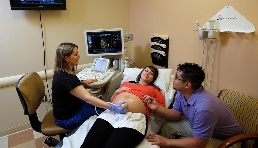
This kind of imaging is otherwise called sonography. Ultrasound utilizes high recurrence sound waves. A gadget called a transducer is situated on the skin over the region of the body part that will be imaged. The transducer discharges sound waves that movement through the skin to the designated organ or tissues. As the sound waves arrive at the particular structure, reverberations are discharged. The transducer will get the reverberations. The reverberations are then changed to a physical image that is shown on a video screen. Ultrasound is utilized in checking a creating embryo, and furthermore in surveying such structures as the kidney, gallbladder, and heart.
- Magnetic Resonance Imaging MRI
This imaging framework utilizes a powerful magnet, radio recurrence signals, as well as a PC which is utilized to make images. It is utilized to analyze spine and cerebrum diseases as well as evaluate joints, mid-region, bone and delicate tissue irregularities, and the chest.
- Atomic Medicine Imaging
This type of imaging utilizes radioactive mixtures that produce gamma rays. The patient ingests a radionuclide. The synthetic compounds collect for a brief time frame in the pieces of the body that are being imaged. A camera detects the gamma rays and uses the information to make an image of the body part. The image is put on film known as a scintigram. It is generally normal utilized in bone and heart appraisal.
The above list features a couple of the various kinds of imaging frameworks utilized in medical diagnosis. Digital Imaging Systems have turned into a fundamental instrument to diagnosing disease and irregularities that outcome in treatment those recoveries lives.